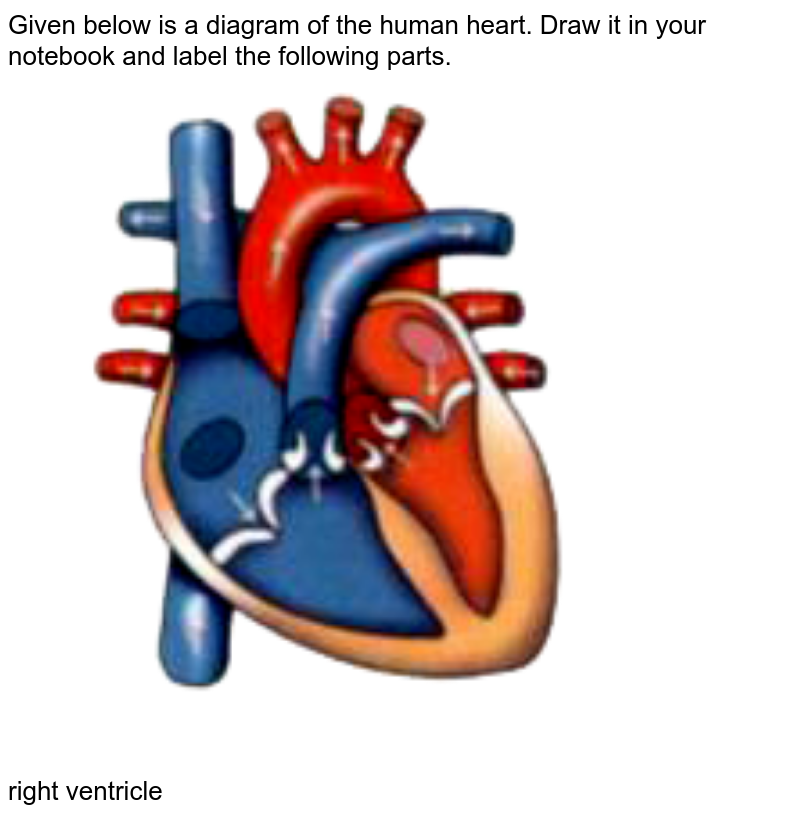
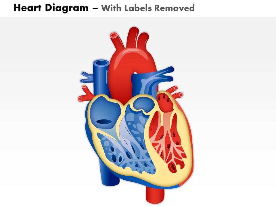
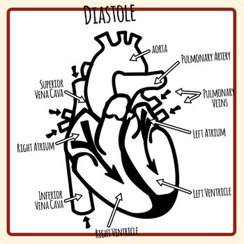
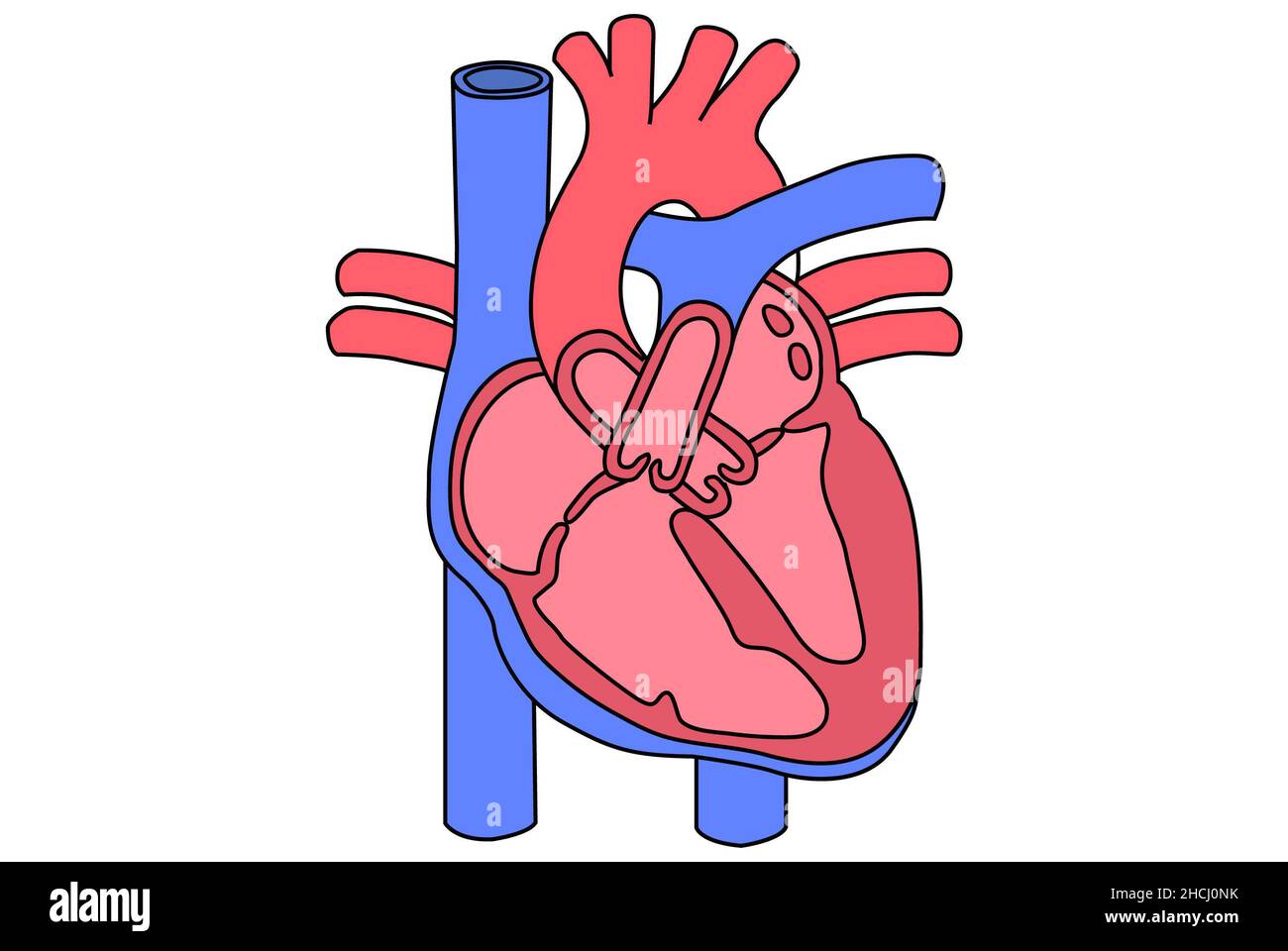
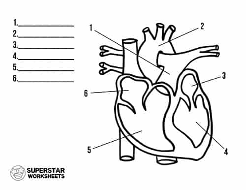
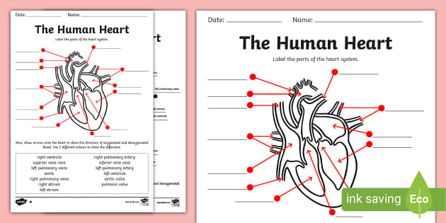
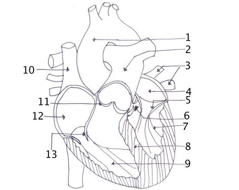
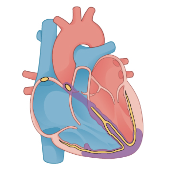
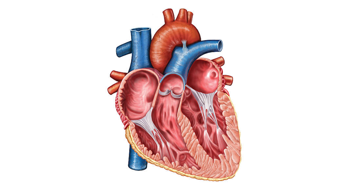
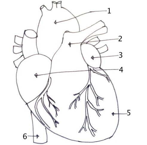
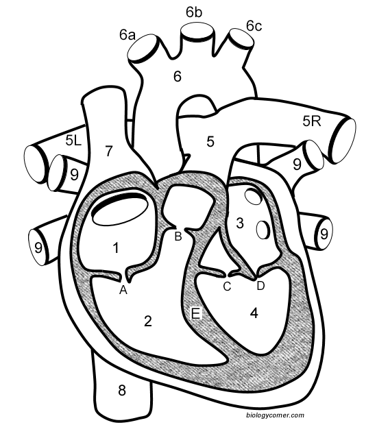
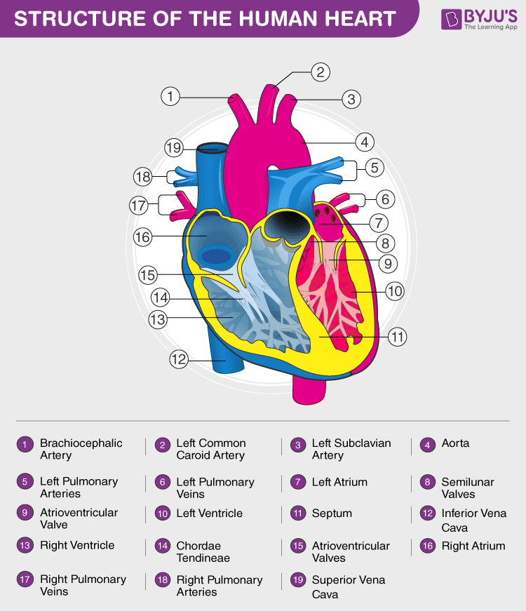
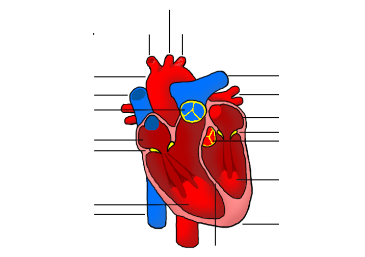
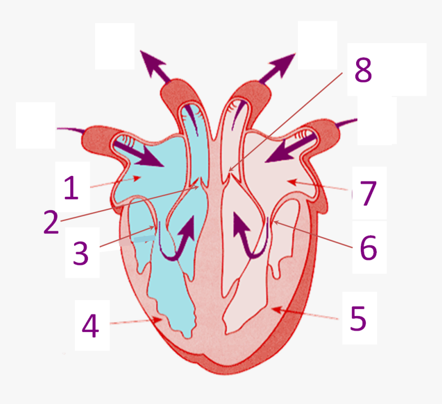
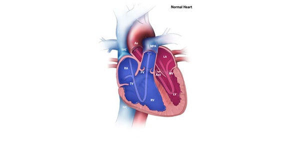
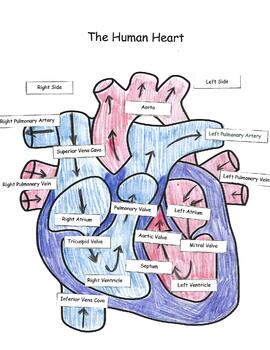
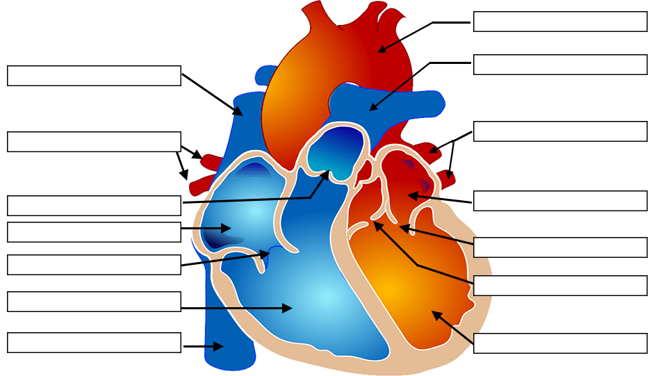
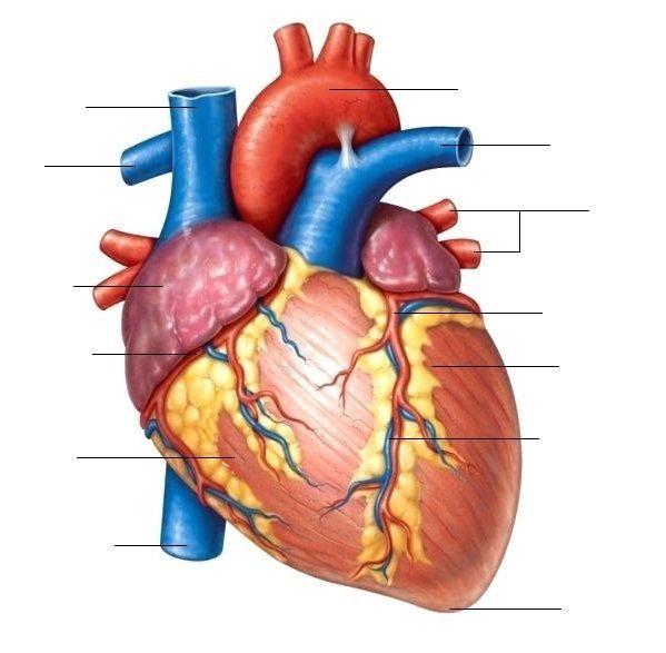
43 diagram of the heart without labels
How the Heart Works - The Heart | NHLBI, NIH - National Institutes of ... The heart is an organ about the size of your fist that pumps blood through your body. It is made up of multiple layers of tissue. Your heart is at the center of your circulatory system. This system is a network of blood vessels, such as arteries, veins, and capillaries, that carries blood to and from all areas of your body. › plot-diagram-template17+ Plot Diagram Template - Free Word, Excel Documents ... From the outside, this plot diagram looks like a network of short and long lines. It is only upon using the template that you will know the effectiveness of the diagram in it. There is an empty field at the top for the title of the story as well. You may also see our circle Venn diagram templates. Printable Plot Pyramid Mountain Diagram Template
How the Heart Works: Diagram, Anatomy, Blood Flow - MedicineNet The heart is located under the rib cage -- 2/3 of it is to the left of your breastbone (sternum) -- and between your lungs and above the diaphragm. The heart is about the size of a closed fist, weighs about 10.5 ounces, and is somewhat cone-shaped. It is covered by a sack termed the pericardium or pericardial sack.
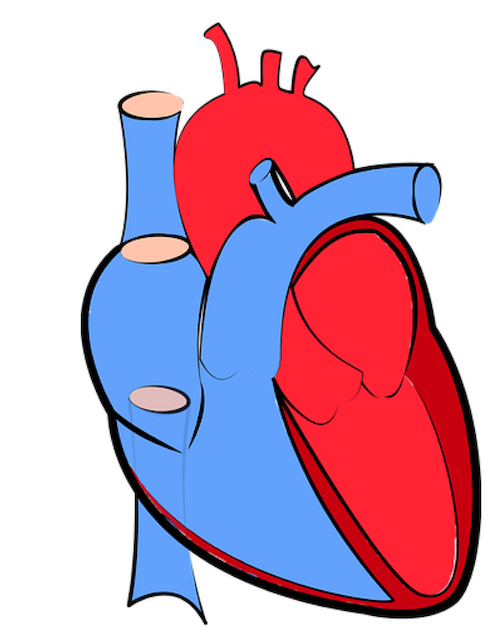
Diagram of the heart without labels
Heart anatomy: Structure, valves, coronary vessels | Kenhub Let's put into words the heart blood flow diagram: Right atrium of heart Atrium dextrum cordis 1/7 The right atrium receives deoxygenated blood from the superior and inferior venae cavae and coronary sinus The right atrium contracts pushing blood through the right atrioventricular valve into the right ventricle. Heart Labeling Quiz: How Much You Know About Heart Labeling? Here is a Heart labeling quiz for you. The human heart is a vital organ for every human. The more healthy your heart is, the longer the chances you have of surviving, so you better take care of it. Take the following quiz to know how much you know about your heart. Questions and Answers. 1. Total Artificial Heart - What Is Total Artificial Heart? | NHLBI, NIH A total artificial heart (TAH) is a pump that is placed in the chest to replace damaged heart ventricles and valves. (Ventricles pump blood to the lungs and other parts of the body.) Once the pump has been placed in the chest, a machine called a driver controls the pump outside the body. The pump and driver help blood flow to and from the heart ...
Diagram of the heart without labels. Human heart: Anatomy, function & facts | Live Science The human heart is located in the center of the chest - slightly to the left of the sternum (breastbone). It sits between your lungs and is encased in a double-walled sac called the pericardium,... en.wikipedia.org › wiki › Yin_and_yangYin and yang - Wikipedia The principle of yin and yang is represented by the Taijitu (literally "Diagram of the Supreme Ultimate"). The term is commonly used to mean the simple "divided circle" form, but may refer to any of several schematic diagrams representing these principles, such as the swastika, common to Hinduism, Buddhism, and Jainism. Cardiovascular system diagrams, quizzes and free worksheets In this diagram of the cardiovascular system, you can see labeled structures. Spend a few minutes analysing the diagram, and trying to connect the location of the structures with what you've learned in the video. Once you think you've got a solid idea, it's time to try our cardiovascular system labeling quiz. Path of Blood Through the Heart | New Health Advisor Here are the basic parts of the heart: 1. Right Atrium The heart can be divided into right and left halves, as well as into the upper and lower chambers. There are two upper chambers called atria and two lower chambers called ventricles. On each side of the heart, there are one atrium and one ventricle.
Circulatory System Diagram | New Health Advisor Coronary circuit mainly consists of cardiac veins including anterior cardiac vein, small vein, middle vein and great (large) cardiac vein. There are different types of circulatory system diagrams; some have labels while others don't. The color blue stands for deoxygenated blood while red stands for blood which is oxygenated. Anatomy of the heart and coronary arteries (coronary CT) - e ... - IMAIOS Anatomy of the human heart and coronaries: how to view anatomical structures. This tool provides access to an MDCT atlas in the 4 usual planes, allowing the user to interactively discover the heart anatomy. The images are labeled, providing an important medical and anatomical tool. The quiz mode makes it possible to evaluate the user's progress. How to Make a DIY Pumping Heart Model | Mombrite Secure with a rubber band or tape. 8. Push both straws through the holes of the balloon. 9. Set the heart model in a tray to catch the "blood.". Make sure to bend the straws downward to avoid projectile blood! 10. Gently press the center of the stretched balloon to pump the blood out of the jar. en.wikipedia.org › wiki › MindMind - Wikipedia Broadly speaking, mental faculties are the various functions of the mind, or things the mind can "do". Thought is a mental act that allows humans to make sense of things in the world, and to represent and interpret them in ways that are significant, or which accord with their needs, attachments, goals, commitments, plans, ends, desires, etc. Thinking involves the symbolic or semiotic mediation ...
› circulatory-system-diagramCirculatory System Diagram - Cardiovascular System and Blood ... They may come with or without labels. Common circulatory system diagrams show pulmonary circulation, coronary circulation, systematic circulation, veins, arteries, or a combination. The systemic circulation system is the most commonly illustrated of the systems that make up the circulatory system as it is the largest. Structure and Function of the Heart - News-Medical.net The heart consists of four chambers, four one-way valves, and a set of arteries and veins that regulate the normal flow of blood within the body. The smooth functioning of the circulatory system ... Heart: illustrated anatomy - e-Anatomy - IMAIOS This interactive atlas of human heart anatomy is based on medical illustrations and cadaver photography. The user can show or hide the anatomical labels which provide a useful tool to create illustrations perfectly adapted for teaching. Anatomy of the heart: anatomical illustrations and structures, 3D model and photographs of dissection jamanetwork.com › journals › jamaoncologyInstructions for Authors | JAMA Oncology | JAMA Network Photographs, clinical images, photomicrographs, gel electrophoresis, and other types that include labels, arrows, or other markers must be submitted in 2 versions: one version with the markers and one without. Provide an explanation for all labels, arrows, or other markers in the figure legend.
Brigitte Zimmer plant and animal cell diagram without labels plant and animal cell drawing easy plant cell and animal cell diagram class 9th plant cell diagram drawing ... simple diagram of a heart with labels simple diagram of a human heart simple diagram of chlamydomonas simple diagram of heart class 10th
Anatomy of The Human Ribs - With Full Gallery Pictures! The Anatomy of the Human Ribs (costae) are one of the integral parts of the chest wall; they make up the lateral part of our body, its anterior and posterior wall and they entirely build the lateral parts of the chest wall. The anatomy of the human ribs is made up of 24 ribs. These ribs are parted in 12 pairs (each on the left and right side of ...
10 Diagram Examples for Any Type of Project - ClickUp 1. Mind map diagram. If you've ever used project management software like ClickUp, you've probably heard of this popular workflow diagram. Mind maps are an excellent tool for planning workflows, visualizing task dependencies, brainstorming new processes —and feeling creative while doing it!
The Location, Size, and Shape of the Heart | GetBodySmart Learn the anatomy of the heart from scratch with these interactive quizzes and free fill-in-the-blank exercises. The narrow end of the heart, which is called the apex, is directed downward and to the left. The apex of the heart. 1 2 It is located at the approximate level of the fifth or sixth rib, just above the arch of the diaphragm.
› blog › heart-anatomy-labeledHeart Anatomy: Labeled Diagram, Structures, Blood Flow ... Feb 24, 2021 · Function and anatomy of the heart made easy using labeled diagrams of cardiac structures and blood flow through the atria, ventricles, valves, aorta, pulmonary arteries veins, superior inferior vena cava, and chambers. Includes an exercise, review worksheet, quiz, and model drawing of an anterior vi
Atrioventricular Node (AV Node): Function and Purpose - Verywell Health The AV node is a tiny "button" of specialized cells (roughly 3 by 5 millimeters in diameter) located near the center of the heart. It is on the right side of the atrial septum at the junction of the atria and the ventricles. 1. Its job is to help coordinate the contraction of the atria and the ventricles in response to the heart's electrical ...
(Solved) - Draw a diagram of the science room and label the locations ... Draw a diagram of the science ...
Ceiling Fan Parts, Names, Functions & Diagram - slidingmotion The electric motor is called the heart of the ceiling fan. Its function is to convert electrical energy into mechanical energy. When an electric current passes in the coil of the electric motor, the magnetic field generates within the coil leads to the coil rotating. This rotation energy is transferred to the fan blades.

Human Heart Circulatory System Diagram Chart Medical Educational Science Class Anatomy Corazon Veins Arteries Labels Black Wood Framed Art Poster ...
Female Anatomy: Labeled Diagrams of the Reproductive System Female anatomy refers to the internal and external structures of the reproductive and urinary systems. Reproductive anatomy aids with sexual pleasure, getting pregnant, and breastfeeding a baby. The urinary system helps rid the body of toxins through urination (peeing). The Female Reproductive System. Some people are born with internal or ...
› ajwThe Asahi Shimbun | Breaking News, Japan News and Analysis Oct 04, 2022 · The Asahi Shimbun is widely regarded for its journalism as the most respected daily newspaper in Japan. The English version offers selected articles from the vernacular Asahi Shimbun, as well as ...
EKG Interpretation & Heart Arrhythmias Cheat Sheet - Nurseslabs Atrial flutter is an abnormal rhythm that occurs in the atria of the heart. Atrial flutter has an atrial rhythm that is regular but has an atrial rate of 250 to 400 beats/minute. It has sawtooth appearance. QRS complexes are uniform in shape but often irregular in rate. Normal atrial rhythm Abnormal atrial rate: 250 to 400 beats/minute
Biblical Counseling Coalition | The Heart Diagram I created this heart diagram to give a very basic explanation of what we mean when we say "target the heart." This is not in-depth teaching; it is a simplistic overview for the sake of helping a counselee understand the process they will undergo in biblical counseling. It is also a tool they can use and apply to specific struggles they may have.
Heart - Wikipedia The heart is a muscular organ in most animals.This organ pumps blood through the blood vessels of the circulatory system. The pumped blood carries oxygen and nutrients to the body, while carrying metabolic waste such as carbon dioxide to the lungs. In humans, the heart is approximately the size of a closed fist and is located between the lungs, in the middle compartment of the chest.
Anatomical Planes of Body | What Are They?, Types & Position In Body Longitudinal plane. A longitudinal plane will be perpendicular to. the transverse plane. It divides the body into two halves and cut the person. straight into left and right halves from the head through the belly button and. down to the toes. The sagittal plane, coronal plane, and parasagittal plane are.
Free Skeletal System Worksheets and Printables - Homeschool Giveaways Skeletal System Worksheets for Kids - These skeletal system worksheets are perfect for younger kids. These sheets will help your kids learn the different bone names. Human Skeleton Printables - This pdf file includes worksheets to review the names of the bones, a fill-in-the-blank page, and a build-your-own-skeleton.
Parts of the Heart & Blood Flow | Diagram & Overview - Video & Lesson ... Diagram of Heart Valves The first valve is the tricuspid valve. It gets its name because it has three cusps that anchor it down into the right ventricle. The tricuspid valve is located at the exit...
heart | Structure, Function, Diagram, Anatomy, & Facts The heart consists of several layers of a tough muscular wall, the myocardium. A thin layer of tissue, the pericardium, covers the outside, and another layer, the endocardium, lines the inside. The heart cavity is divided down the middle into a right and a left heart, which in turn are subdivided into two chambers.
Chest (lateral view) | Radiology Reference Article - Radiopaedia Otherwise, a left lateral view is the default and preferred side as it demonstrates better anatomical detail of the heart. Patient position. standing upright; left side of the thorax adjacent to the image receptor. left shoulder placed firmly against the image receptor; both arms raised above the head, preventing superimposition over the chest
Total Artificial Heart - What Is Total Artificial Heart? | NHLBI, NIH A total artificial heart (TAH) is a pump that is placed in the chest to replace damaged heart ventricles and valves. (Ventricles pump blood to the lungs and other parts of the body.) Once the pump has been placed in the chest, a machine called a driver controls the pump outside the body. The pump and driver help blood flow to and from the heart ...
Heart Labeling Quiz: How Much You Know About Heart Labeling? Here is a Heart labeling quiz for you. The human heart is a vital organ for every human. The more healthy your heart is, the longer the chances you have of surviving, so you better take care of it. Take the following quiz to know how much you know about your heart. Questions and Answers. 1.
Heart anatomy: Structure, valves, coronary vessels | Kenhub Let's put into words the heart blood flow diagram: Right atrium of heart Atrium dextrum cordis 1/7 The right atrium receives deoxygenated blood from the superior and inferior venae cavae and coronary sinus The right atrium contracts pushing blood through the right atrioventricular valve into the right ventricle.

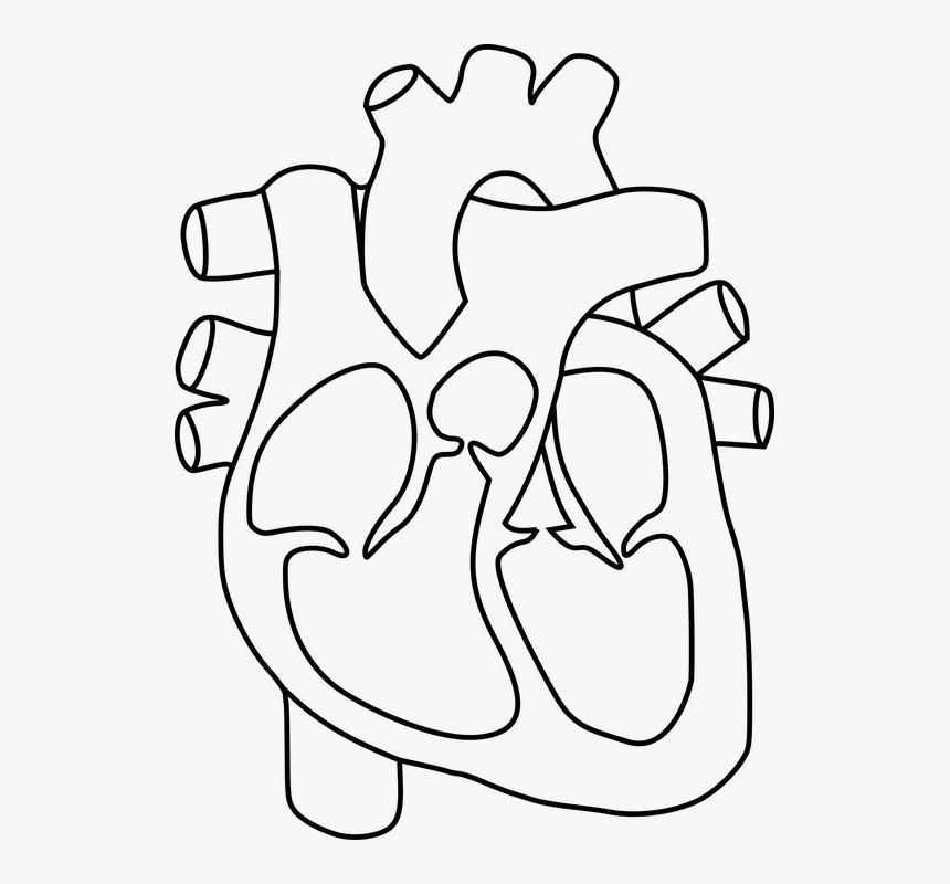
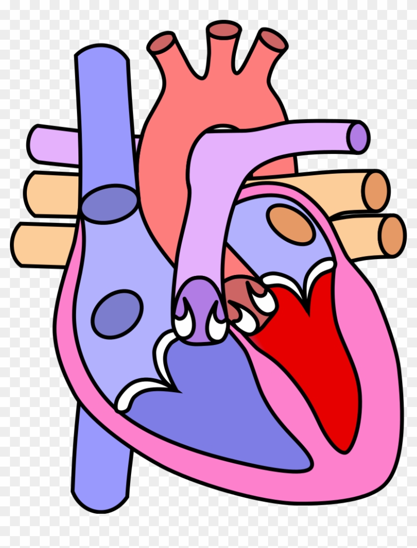









Post a Comment for "43 diagram of the heart without labels"